
Your care pathway
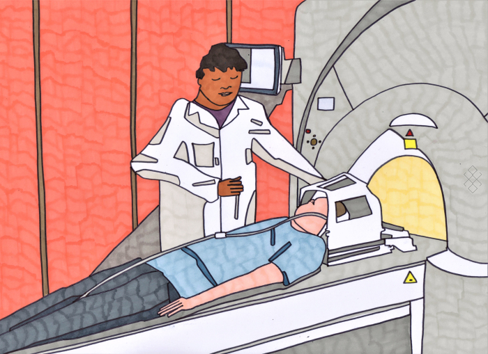
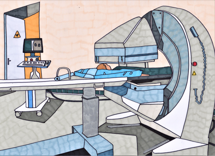
MRI
MRI uses electromagnetic waves with a large magnet to construct images in two or three dimensions. It allows viewing of specific elements of the body using the water protons.
Read more ⟶
Mammography
A mammogram is a breast X-ray. It uses low-dose X-rays and provides breast cancer screening.
Read more ⟶
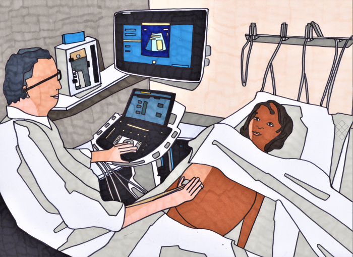
Ultrasound & Doppler
Ultrasound uses ultrasonic waves to construct the image of the different organs of the body. Using a probe which sends an ultrasound beam, images are obtained in real time.
Read more ⟶
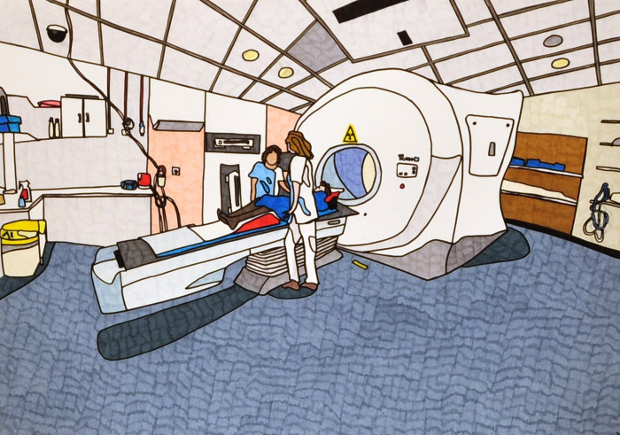
Scanner
CT uses X-rays with a tomographic analysis technique to produce images in thin sections. Reconstruction of tissue scans can be done in 2 or 3 dimensions.
Read more ⟶
Medical Imaging
All medical imaging examinations
Read more ⟶
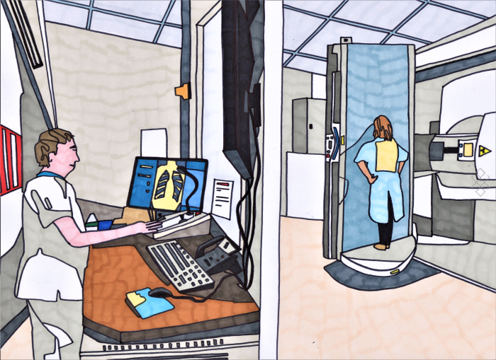
Conventional Radiology
Radiography uses X-rays through the body to image gray scale in two dimensions. The absorption of X-rays depends on the density of the organ X-rayed.
Read more ⟶
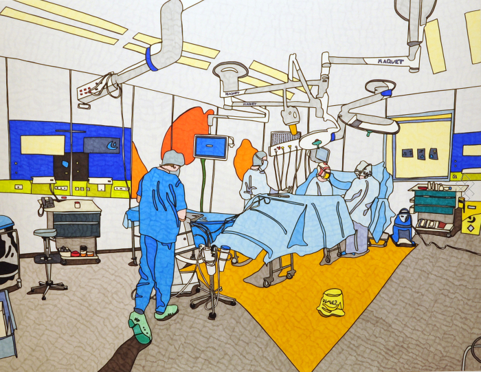
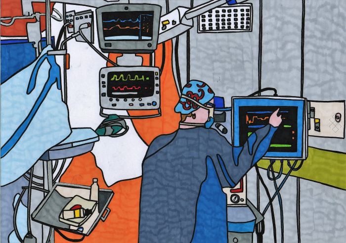
Interventional Radiology
Interventional radiology allows for therapeutic or diagnostic actions in different imaging techniques, with the most common being the use of X-rays.
Read more ⟶
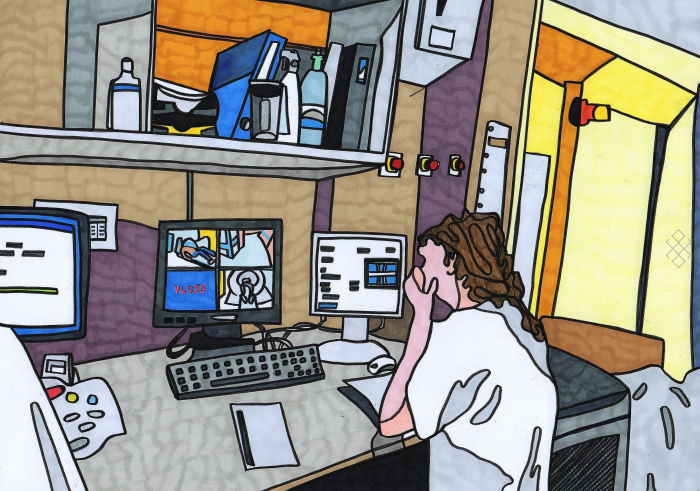
Scintigraphy
The scan uses radioactive tracer that attaches to organs. Analysis of cellular uptake allows for two dimensions (scintigraphy) or three dimensions images (PET-CT).
Read more ⟶
Endoscopy
Endoscopy or fibroscopy to visualize the interior of hollow organs. Through the introduction of a camera into a hole it allows for a diagnostic or therapeutic procedure.
Read more ⟶
Partners and supporters


Legal notice - Politique de Protection des Données - CGU
Copyright © 2020 - 2026 Alphath Access ltd. All rights reserved